Project Research
Vesalius Trust Research Scholarship Winner
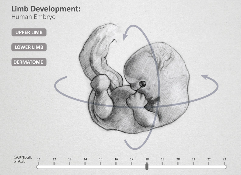
Visualizing the Development of Upper and Lower Limbs;
Formation of Dermatome during Human Development: Weeks 4-8
Goals and Objectives
The goal of this research is to improve students’ understanding of upper and lower limb development via a 3D interactive program, to allow a more thorough understanding of how flexion and extension occur in different planes in the adult limbs. In addition, this interactive program may also improve students’ understanding of the relationships between those bending and rotational movements and the origin of the dermatome patterns found in adult anatomy. With a more complete visual understanding of limb development, dermatome maps and the cutaneous nerve distribution in adult limbs, medical students may be more successful in clinical diagnosis in the future.
Audience
The intended audience is first year medical students. The interactive 3D program was created for first year medical students enrolled in BMS 645 Medical Gross Human Anatomy and Embryology II at the University of Illinois at Chicago (UIC) College of Medicine (COM).
Materials and Methods
The Digitally Reproduced Embryonic Morphology (DREM) data was used to digitally reconstruct 3D models for reference and to ensure accuracy of the project3. The cross-sections of embryos in different embryonic stages from DREM data set was brought into Materialise Mimics. Edited embryo models were exported and optimized in Autodesk 3ds Max. The 3D models and animation of upper and lower limb morphology were created using Autodesk 3ds Max, Pixologic Z-Brush and The Foundry MODO. 3D animation was used to present the upper and lower limb morphology with its corresponding cutaneous nerve innervation. Unity 3D and JavaScript was used to create the interactive user interface. The finished 3D interactive tool was consist of a 3D models and interactive 3D animation.
Project Concept
Rapid and accurate learning of human embryology by first year medical students informs normal adult structures, normal anatomical variations, congenital anomalies and supports clinical diagnosis1. Visualizing the 3-dimensional changes that include complex and unintuitive bending and rotation during limb development is especially difficult. The limb growth process doesn’t parallel adult limb orientation. In addition, students are expected to understand dermatome development, as it directly relates to the bending and rotation that occurs during limb growth and development 2.
Currently, there are many 2D illustrations representing the development of the dermatome maps. However, students struggle to grasp all of the rapid changes in embryonic limb and dermatome development together 3. Understanding embryonic limb development correctly from the beginning provide medical students with a robust and reliable foundation for conceptualization of anatomical relationships in the limbs. Visualization of this process leads to the formation of a mental image that is critical to providing directional orientation and context. The sequence of events during limb development will need to be recalled repeatedly in the clinical setting in order to correctly diagnose and treat patients with birth defects or injuries to the limbs.
Under the supervision of Embryologist and UIC College of Medicine Medical Gross Anatomy & Embryology course director Dr. Maurice Pescitelli, this research project developed an interactive 3D program. This 3D interactive tool depicted the bending and rotation of upper and lower limb development, in addition to visualizing dermatome map migration and its corresponding nerve distribution.
Preproduction

A visual growth chart of human embryos from Carnegie stages 12 to 23 was created as a visual reference for 3D model building. This chart consisted of references images and photographs obtained from the Visible Human Embryo Project (VHE) website and University of New South Wales (UNSW) Embryology online education and research website
Production


Production Image
References
1 Carlson, B., (2002) Embryology in the medical curriculum. The Anatomical Record. 269 (2002)
89–98.
2 Moore, K. L. & Persaud, T.V.N. (2008) The Limbs (8th). The Developing Human. Philadelphia,
PA: Saunders Elsevier.
3 Larsen, W.J. (2001). Human Embryology: Third Edition. PA: Churchill Livingstone.